RESEARCH
Neuropeptide regulation of thalamocortical networks in sleep and arousal
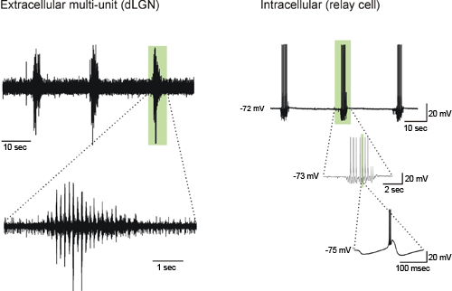
Neurones in higher brain regions such as the thalamus and cerebral cortex display strikingly different patterns of activity when we are awake and when we sleep. Our work focuses on determining the role of neuropeptides in shifting the network between the slow oscillations typical of non-REM sleep and the tonic firing conducive to sensory processing in the awake state. For this purpose, we make use of unique slice preparations of the sleeping forebrain developed by David McCormick and collaborators at Yale University (von Krosigk et al., 1993; Sanchez-Vives and McCormick, 2000). In this model, we investigate network activity patterns with extracellular recordings and underlying cellular mechanisms with intracellular and patch clamp recordings. Previous work (Broberger and McCormick, 2005) has e.g. demonstrated that the neuropeptide thyrotropin-releasing hormone (TRH) exerts powerful excitatory actions within the thalamic network, providing a mechanism for the arousal-promoting and anti-epileptic actions of this peptide shown clinically and in experimental studies. - We are also interested in how cortical activity is modulated by input from the lateral hypothalamic area mediated via the neuropeptides hypocretin (orexin) and melanin-concentrating hormone. These peptides are implicated in the control of sleep and arousal. Most notably, an almost complete lack of hypocretin-expressing neurones has been described in narcoleptic patients.
Brain circuitries regulating food intake and appetite
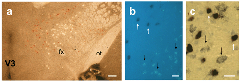
The brain is responsible for regulating energy metabolism and, as a result, body weight. To accomplish this task, the central nervous system needs to inform itself about the metabolic state of the body, which it does via two main channels. In the arcuate nucleus of the hypothalamus, metabolically relevant hormones such as leptin and insulin relay information about the supply and demand of energy in the body, and act on two antagonistic populations of neurones: feeding-stimulatory neurones expressing neuropeptide Y (NPY) and feeding-inhibitory neurones expressing pro-opiomelanocortin (POMC). In the brainstem, the vagus nerve transmits sensory signals from the gastrointestinal tract in the nucleus tractus solitarii (nTS). In previous work, we have studied the ascending projections from the arcuate nucleus and found that NPY- and POMC-ergic neurones project in pathways parallel to each other and to cells originating in the nTS (e.g. Broberger et al., 1998). We are interested in defining the target neurones for this triple innervation to understand how higher brain regions are engaged in the initiation, organization and termination of feeding behaviour. We have also studied the hypothalamic histochemistry in a genetic model of fatal anorexia (the anx/anx mouse), and found changes centred in the arcuate nucleus, which may underlie the failure-to-thrive in this mutant model (Broberger et al., 1997; 1998; 1999).
Broberger C. and McCormick DA, 2005. Excitatory effects of thyrotropin-releasing hormone (TRH) in the thalamus. J. NEUROSCI.; 25(7): 1664-1673
Broberger C. et al., 1999. Changes in neuropeptide Y receptors and pro-opiomelanocortin in the anorexia (anx/anx) mouse hypothalamus. J. NEUROSCI., 19 (16): 7130-7139
Broberger C. et al., 1998. The neuropeptide Y agouti gene-related protein (AGRP) brain circuitry in normal, anorectic, and monosodium glutamate-treated mice. PROC. NAT. ACAD. SCI. USA, 95 (25): 15043-15048
Broberger C et al., 1997. Hypothalamic neurohistochemistry of the murine anorexia (anx/anx) mutation: Altered processing of neuropeptide Y in the arcuate nucleus. J. COMP. NEUROL., 387 (1): 124-135
Sanchez-Vives M.V. and McCormick D.A., 2000. Cellular and network mechanisms of rhythmic recurrent activity in neocortex. NATURE NEUROSCI., 3:1027-34
von Krosigk M. et al., 1993. Cellular mechanisms of a synchronized oscillation in the thalamus. SCIENCE, 261:361-4